Trepanation, or trephining, may well represent the earliest wound inflicted by one human being on another for the purpose of healing and can be considered the beginning of the practice of surgery and the earliest tangible evidence of medical practice (1). In trepanation, a portion of the skull is removed. Some of the oldest examples of skulls showing trepanation defects, from Peru and other sites, demonstrate holes at or near a recent crack and most likely represent an attempt to relieve the effects of a skull fracture (1-4). 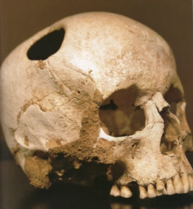 Many defects show evidence of healing, indicating that the procedure, even thousands of years ago, was not necessarily fatal.
The fact that many trepanated skulls have been recovered indicates that the procedure was often therapeutically successful and repeatedly applied. Indeed, differences in technique can even be discerned, particularly among the Incas who were masters in the art of trepaning (5). It may even be that bone “transplants” were used to fill in defects (6).
The first trepanation may have taken place as long as 10,000 years ago (1, 7-10). Examples of 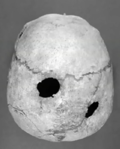 trepanation have been found in many parts of the world, including multiple sites in Africa, Asia, Europe, North and South American, Central America and Melanesia (1, 7, 10-19).
In addition to its clear therapeutic indication for trauma to the skull, trepanation may have been used as a ritual or magical rite, perhaps, as many authorities have suggested, to cast out demons thought to have taken possession of an individual (1, 10, 12). This theory is supported by the failure to find any sign of prior cranial injury in many of the skulls from the neolithic period, when stone carving and tool creation began. Those hapless individuals, labeled as being inhabited by the devil, might have suffered from epilepsy, migraine headaches, psychologic and psychotic disturbances, and a variety of cerebral organic disorders, including brain tumors. The ancient Pharaohs underwent trepanation shortly before death to allow their souls to leave their mortal bodies and go up to heaven (20).
Those unknown ancient surgeons, as they should appropriately be regarded, likely had considerable skill and great courage. There is no information about how the patient submitted to such an operation. Was there some powerful opiate available? Sedation with herbs was known during the Han dynasty in China (14). Did the patient submit voluntarily, observing his fellow tribesmen incise his scalp, cut the bone and open the skull? Did a number of strong men hold the patient down? A variety of trepanation techniques have been demonstrated (17).
In comparatively recent times, Melanesians performed trepanation for headache, vertigo, and epilepsy, as well as for cranial injuries. In his work On Wounds in the Head, Hippocrates describes his use of trepanation, including a fairly detailed description of how to perform the operation (21), cautioning the operator to avoid penetrating the skull too quickly and thereby injuring the dura mater (22), the covering of the brain. Celsus also described trepanation for the relief of head wounds (22). Over the centuries, trepanation has been used for “practically everything related to the head” (11), including stammering (23). In the Medieval period, Lanfranc, a student of Saliceto who was originally known as Lanfranchi of Milan, and who was the first to describe concussion of the brain and the classic signs of skull fracture, used trepanation to relieve depressed skull fractures but also for “irritation of the dura” (24) which is not better 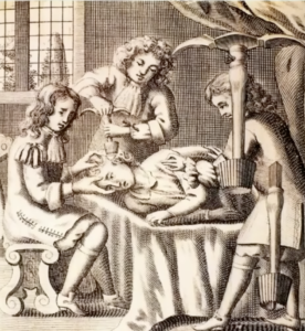 defined. Woodcuts from the Renaissance era who trepanation being performed.
  The great Dutch painter, Heironymus Bosch (1450-1510) showed the operation in his “The Cure for Madness.”
In our time, of course, trepanation, or craniotomy, is no longer utilized to treat epilepsy, dementia, or psychosis, although it was not too long ago that craniotomy or a less invasive modification of craniotomy, passing probes into the brain via a transorbital approach (“prefrontal lobotomy”), was employed to treat manic behavior. The Ken Keysey novel, and subsequent film, One Flew Over the Cuckoo’s Nest, depict this vividly.
Trepanation derives from the Greek word trema, which is synonymous to trupa, from which trupan, to pierce or bore, derives (25). From there comes trupanon, a piercing or boring instrument, the Medieval Latin trupanum, a surgical instrument, as well as the Medieval French trépane, and its derivatives trépan, trépaner and trépanation. The English trepan, “to trepan,” and trepanation became modified to trephine, an improvement on the trepan, “to trephine,” and trephination. 17th Century Trepanation 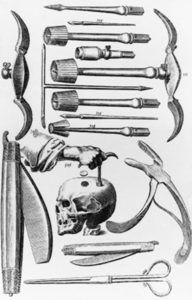 kits, including all the necessary instruments, used by the British Navy were illustrated.
It is of some interest that trepanation under fairly primitive conditions has always been a relatively safe procedure, with a survival rate approaching 100 percent (1, 4, 6, 7,10). Although there is almost no documentation of the ways in which wounds were treated before Hippocrates, the majority of recovered skulls show good evidence of long-term healing of the skull defect. There is evidence, in different civilizations, of different techniques of trepanation (1, 5, 6), but there is little record of the types of poultices and salves that must have been used to combat infection until Hippocrates described a form of tar that he used on wounds (18, 26).
In contrast the this history of safe performance, the same operation, during the first half of the 19th century, had a mortality rate that also approached 100 percent. In this period, trepanation was regarded as so dangerous that the first requirement for the operation, wrote one authority, was “dass der Wundarzt selbst auf den Kopf gefallen sein müsse” – “that the wound surgeon himself must have fallen on his head” (1). During this period, however, a Kentucky surgeon, Benjamin Dudley, performed trepanation on six patients with post-traumatic epilepsy in the years 1819 to 1832. He considered it a safe operation, having learned the technique in Paris from Larrey, Dupuytren and other French surgeons (27).
One of the first to recognize that the cranial defects of trepanation were the result of operations performed for therapeutic purposes was Paul Broca, one of the pioneer neurologists and founder of modern neurosurgery, as well as a professor of surgical pathology at the University of Paris, in 1876 (24, 26, 28). Broca was also an anthropologist. His studies contradicted the conclusions of Prunières, a physician interested in archaeology, who, after finding a skull with a large occipital opening with smooth edges in an ancient tomb in central France, had concluded that the skull had been perforated for use as a drinking cup. Broca discovered the speech center of the brain, which led to the mapping out of the various functional centers of the brain. Indeed, Broca was the first to perform trepanation at a specific site he identified on the basis of his theory of localization of function (24).
Trepanations were also performed after death for ritual purposes. It is thought that the ancient Egyptians did this on many bodies, in addition to Pharoahs, in order to extract the brain so that the head would more easily undergo embalming to assure preservation of the body as perfectly as possible for “the life after death” (21). Trepanation after death may also have been performed to obtain small portions of bone to be used as amulets (5, 17).
Self-trepanation is well-documented and performed, ostensibly, for therapeutic reasons (29, 30), as well because of psychiatric illness (31). Indeed, one Englishwoman, who performed trepanation on herself, 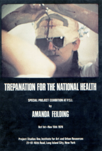 campaigned unsuccessfully for a seat in Parliament, including as one of her campaign proposals that trepanation be made freely available as a part of the National Health Service programs (29). There are many discussions of trepanation on the internet (32).
But this blog posting is not solely for the purpose of reviewing the fascinating history of trepanation. Instead, it is to provide first-hand confirmation of the adage that a physician who treats himself/herself has a fool for a patient.
My trepanation, or craniotomy, occurred on May 21, 2000, for relief of a fairly sizable “subacute” subdural hematoma.
My subdural hematoma most likely resulted from a sharp blow to my head, approximately five to six weeks before, sustained while installing a new computer under my desk at home. This event was only recalled when I was hospitalized. Modern computer desks are generally fairly light and easily movable, and probably would not cause any significant injury under these circumstances. My desk, which I have had since I was in junior high school, in Brooklyn, New York, in 1950, is solid oak and literally requires two strong men to move from one site to another. There were no immediately recognizable effects. I did not have so much as a “bump” at the site.
Approximately four weeks prior to the craniotomy I started developing headaches which became increasingly persistent and severe. I have been most fortunate in my life and have almost never had headaches, despite a strong family history of migraine headaches. The last time I had a headache requiring aspirin had been five or more years before. At that time I had a mid-50’s attack of guilt at not exercising regularly and did a series of pushups, following which I had a severe headache that spontaneously resolved in a day or so. I happened to pass one of my colleagues, an outstanding neurologist, in the hospital corridors and described this event. I asked him whether it might be the earliest sign of an aneurysm of some other untoward vascular event. He assured me that it was probably just from the exercise and nothing to worry about but, if I was really concerned, “it is easy enough to get an MRI.” Of course, at the time, this recommendation might not have been tenable in every community, but Beverly Hills, with its population of approximately 40,000 people, reputedly had more MRI instruments than in all of Canada, with is population of more than 25,000,000 people. A renowned neuroradiologist, Chairman of the Department of Imaging, assured me that the MRI which I decided to avail myself of, including the cerebral vessels, was 100% normal.
I was traveling each of the three weekends before my craniotomy. I was in Chicago for a meeting. Then, the first weekend in May, I was delightfully spent in San José, California, attending attending a performance of La Traviata by the San José Opera Company – conducted by my friend and fellow pathologist, Dr. William J. Siegel. It was one of the best performances I had ever heard but I was also starting to use analgesics of various kinds on a more regular basis. I even had an exacerbation of my now constant headache when I stepped on and off the sidewalk while crossing a street. The next weekend I set out for Boston, to attend my nephew’s bar mitzvah. This, in retrospect, was probably the scariest part of my entire experience. I left very early in the morning on Friday, May 12 on a continuation flight through Chicago. Unfortunately, this was the weekend of a major “slowdown” by United Airlines pilots as well as the time of heavy Spring storms in the Midwest. I never made it to the bar mitzvah, spending the Friday night in a hotel approximately 10 miles from Ohare Airport, alone and ingesting pain medication on a fairly regularly basis.
I had, a week or so before this, met my neurologist friend again and told him I was having headaches. He peered into my eyes and saw that my pupils were reactive and equal, and that I had no obvious gait disturbances, and suggested that I call him if it continued. He also noted that I had slight left-sided ptosis (drooping upper eyelid), and commented “but you’ve had that for while.”
A few years ago I had unilateral, right-sided ptosis which my then secretary correctly diagnosed as the result of an insect bite and which appropriately resolved spontaneously after a few days. When I returned to my office and looked in the mirror, I remembered this episode and also recognized that the left-sided ptosis was new. However, I did not take the time to ask myself what it meant, being very occupied with work and being annoyed at my headaches.
And continuing to practice my skills in the art of denial.
A few days before my craniotomy the headaches became more prominent frontally. Indeed, any unusual motion of my head, such as shaking my head or stepping down from the sidewalk, much more prominent than it had been, now elicited increasingly severe pain. I now encountered, in a hospital coffee shop, another friend and colleague, an otorhinolaryngologist (ENT), who I had last seen me 15 years before for tinnitus (ringing in the years most like due to years of taking aspirin, to be discussed again below). Since, I had a little nasal congestion I wondered if my headaches could be from sinusitis. I asked him if he would take a look at me, which he did. After the briefest of examinations, late on the Friday afternoon, he was unequivocal that it was not sinusitis and, with a somewhat worried look, sent me for the MRI.
After the MRI procedure I went back to my office. About thirty minutes letter, the chairman of Imaging called to see how I was and invited me to come to the Imaging department suite, just down the hall from my office, to view the results. I was greeted by three neuroradiologists who came out to escort me to the viewing screens. In retrospect, they apparently wanted to confirm with their own eyes that I was able to walk. My first thought, of course, was that I had the most awful brain tumor every recorded. Instead, fortunately, they showed my MRI with a large subdural hematoma (solid white in the image) 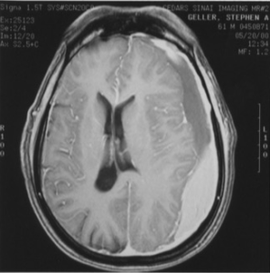 considerably compressing my brain. They then called the neurologist who scheduled me for admission the following morning. One of the three neuroradiologists thought I might want to get second opinion before undergoing surgery, since the subdural had been there for a period of time, but even I could see from the MRIs that it was time to take action.
Since, at that time, my old desk had not yet been recalled as the probable cause, the reason for the bleed was unknown and, prior to surgery, we needed to determine if I had a coagulopathy (disorder of blood clotting). Virtually every aspect of the coagulation cascade was studied, from the standard prothrombin time and platelet aggregometry analysis to analysis of ristocetin co-factor and arachidonic acid and factors VIII, IX, XI, XII and XIII. An abnormality was not identified. All of my other test values, including standard chemistry assays, hematology studies, liver activity evaluations and antinuclear antibody determination were all remarkably unremarkable. With my unremarkable physical examination (at least below the neck), my unremarkable chest x-ray and electrocardiogram, and my long list of unremarkable laboratory test results, I was in remarkably good health as I was about to have my skull opened.
The next morning I was admitted to my then hospital, Cedars-Sinai Medical Center. The unit was on the same floor as my office and I tried to work for as long as possible. I was, however, not highly functional. I managed to disperse my uncompleted cases to various colleague’s mail slots and sent my last e-mail before surgery at about 9:30 PM, informing my colleagues and key hospital personnel about my scheduled Sunday morning procedure.
My neurosurgeon who is professionally exceedingly concentrated and brusque, and with whom I have never been particularly friendly, was, as my caretaker, the model of the best traditions of medicine. I knew well that he was exceedingly competent, but he proved to be completely supportive and informative, caring and patient, and even charming. In order to assure the complete removal of the hematoma, including all portions of the chronic membrane that had formed, he elected to remove, and then replace, a “flap” of cranial bone, approximately 3 x 4 cm. He determined that “burr” (drill) holes would not be sufficient. With the outstanding nursing care I received, and the care and encouragement shown me by a whole range of hospital employees and my colleagues, I did very well. There is no evidence of my adventure other than an 8-10 cm incision on my slightly hairless scalp and a slight depression marking the borders of the flap. Looking back, it is clear that, in addition to my headaches, I was becoming increasingly fatigued, a feeling I ascribed to how busy I was, and I was also just beginning to mix up words, although apparently not obviously to anyone but me and, even by me, fully acknowledged only in hindsight.
Were there contributing factors other than the inertia of my junior high school desk? It is likely that the more than 20 years of aspirin I had taken daily contributed (33).
Was a lesson learned?
I had not had a regular physician since I was in high school, and that was for the purpose of “checkups.” I had a chest x-ray some years ago, when my hospital required it of all physicians on staff but had not had an electrocardiogram for at least 20 years. I have always felt healthy. For screening, I content myself with donating blood on a regular basis, thereby having hemoglobin determination, hepatitis and HIV testing, as well as blood pressure evaluation. In all these regards, as well as annual PSA determination and biannual lipid determination, I was “normal.”
As a part of being admitted, in the highly specialized milieu in which I practiced, I needed a general internist. I offered the name of another friend. When he visited me to take my history, he asked, among other things, whether I had ever had a colonoscopy. I recalled that my hemoglobin and hematocrit determinations are always at very healthy levels and that I had, at least until the present illness, not been fatigued. I also told him that I had no family history of polyps or colon cancer, and probably would not undergo colonoscopy. He suggested I at least think it over, and certainly have my stool tested for occult blood.
When my craniotomy incision, and the rest of me, healed completely, thoughts about that colonoscopy faded for another five years.
References:
- Majno G. The Healing Hand – Man and Wound in the Ancient World. Cambridge, Harvard University Press, 1975, pp 24-26.
- Steward TD. Some problems in human paleopathology, In Jarcho S, Human Paleopathology, New Haven, Yale University Press, 1966, pp 43-55.
- Froeschner EH. Two examples of ancient skull surgery. Journal of Neurosurgery 1992; 76:550-552.
- Gerstzen PC, Gerstzen E, Allison MJ. Diseases of the skull in pre-Columbian South American mummies. Neurosurgery 1998; 42:1145-1151.
- Stewart TD. Pseudotrephination. American Journal of Physical Anthropology 1971; 35:296-297.
- Henschen, Folke. The History and Geography of Diseases. New York, Delacorte Press, 1966, p 251.
- Hershkovitz I, Levi B, Hiss J, Arensburg B. Medicoritual trephinations in modern Israel. Am Journal of Forensic Medical Pathology 1991; 12:194-199.
- Alt KW, Jeunessse C, Buitrago-Tellez CH, et al. Evidence for stone age cranial surgery. Nature 1997; 387:360.
- Lillie MC. Cranial surgery dating back to Mesolithic. Nature 1998; 391:854.
- Lisowski FP. Prehistoric and early history of trepanation. www.trepanation.com/master12.htm.
- Margetts EL. Trepanation of the skull by the medicine-men of primitive cultures, with particular reference to present-day native East African practice, In Brothwell D, Sandison AD, Diseases in Antiquity, Springfield, CC Thomas, 1967, pp 673-701.
- Prioreschi P. Possible reasons for Neolithic skull trephining. Perspectives in Biology and Medicine 1991; 34:296-303.
- Velasco-Suarez M, Bautista Martinez J, Garcia Oliveros R, Weinstein PR. Archaeological origin of cranial surgery: trephination in Mexico. Neurosurgery 1992; 31:313-318.
- Chan EL, Ahmed TM, Wang M, Chan JC. History of medicine and nephrology in Asia. American Journal of Nephrology 1994; 14:295-301.
- Rawlings CE 3rd, Rossitch E Jr. The history of trephination in Africa with a discussion of its current status and continuing practice. Surgical Neurology 1994; 41:507-513.
- Richards GD. Brief communication: earliest cranial surgery in North America. American Journal of Physical Anthropology 1995; 98:203-209.
- Arnott R. Surgical practice in the prehistoric Aegean. Medizinhistory Journal 1997; 32:249-278.
- Sanan A, Haines SJ. Repairing holes in the head: a history of cranioplasty. Neurosurgery 1997; 40:588-603.
- Pick J, Lidke G, Terberger T, von Smekal U, Gaab MR. Stone age surgery in Mecklenburg-Vorpommern: a systematic study. Neurosurgery 1999: 45:147-151.
- Bailey P. Anecdotes from the history of trephining. 1961. Surgical Neurology 1994; 42:83-90.
- Zivanovic S. Ancient Diseases – the Elements of Paleopathology. New York, Pica Press, 1982’ pp 185-191.
- Castiglioni A. A History of Medicine, 2nd ed. New York, Alfred A. Knopf, 1947, pp 26-27, 171, 210.
- Porter R. The Greatest Benefit to Mankind. New York, WW Norton, 1998, pp 35.
- Garrison FH. An Introduction to the History of Medicine, 4th ed.. Philadelphia, WB Saunders, 1929, pp 155, 159, 492.
- Partridge E. Origins – A Short Etymological Dictionary of Modern English. New York, Greenwich House, 1983, p 718.
- Guthrie D. A History of Medicine. London, Thomas Nelson, 1958, pp 6, 11.
- Jensen RL, Stone JL. Benjamin Winslow Dudley and early American trephination for posttraumatic epilepsy. Neurosurgery 1997; 41:263-268.
- Anon. Paul Pierre Broca. www.epub.org.br/cm/n02/historia/broca.htm.
- Mellen J. Hole in the head. The Auger, www.noah.org/trepan/hole in the head.html.
- Michell JF. The people with holes in the heads. The Auger, www.noah.org/trepan/people with holes in their heads.html
- Wadley JP, Smith GT, Shieff C. Self-trephination of the skull with an electric power drill. British Journal of Neurosurgery 1997; 11:156-158..
- Anon. www.fringeware.com/subcult/Trepanation.html
- Reymond MA, Marbet G, Radü EW, Gratzl O. Aspirin as a risk factor for hemorrhage in patients with head injuries. Neurosurgical Review 1992; 15:21-25.
October 21, 2014 at 7:00 pm
fascinating story.
October 21, 2014 at 11:01 pm
Excellent account. Quite similar to my recent experience. On my return from Nigeria on July 17, 2014 (my birthday) I developed an headache for which I had to ask one of the flight attendants for an analgesic. It abated somewhat bur was persistent for another two days. Despite my wife’s request for me to go to the ER, I refused until the evening of July 20th when I had an episode of projectile vomiting followed by dizziness. I then went to the ER where a CT scan showed a subdural hematoma. I was transferred to Westchester Medical Center (NY Med College) where I had a bore hole done and 150cc of blood drained over the next four days. My headache continued for another three weeks after discharge and it took me another few weeks to fully recover. I am now almost back to my old self. I didn’t have any history of significant trauma or a coagulopathy. The neurosurgeons thought that I might have sustained the injury during my traveling on bad roads in Nigeria (untold number of potholes some which I describe as “ditches”). He said one could get a subdural hematoma from a serious whiplash.
Many thanks for sharing this interesting piece with us. I thoroughly enjoyed the history (your forte), your usual succinct and clear style and the way you tied it with your personal experience with this age-old surgical procedure.
I trust all is well with you. I am still trying to obtain your new book as an e-book from the Kindle bookstore.
Segun
October 23, 2014 at 12:57 pm
Segun,
So nice to hear from you. We will have to compare our holes in the head – although yours is probably just a fingertip indentation whereas mine is a depressed plateau.
Not sure why you are having trouble with getting a Kindle copy – I must confess I am not yet fond of electronic reading. Should I send you a print version?
Best wishes.
Stephen
January 27, 2015 at 10:55 pm
I am distantly related to Dr.David Sachar (gastroenterologist) by marriage. His older brother ,Howard Sachar is here in L.A. visiting me. Howard is the famous historian and he brought his 17th book as a gift for us. I remembered that you knew and praised David Sachar. I have many copies of your novel.I gave one to Howard who sees David periodically. Howard will give your novel to David or will mail it to him. I am sure that David will be happy to see what his old friend (you) have written. I think your blog is fascinating. Keep up the blogs. Mort Klein
February 23, 2015 at 5:36 pm
Mort,
You are my unofficial agent!
I recently received a lovely note from David Sachar who I know for a long, long time. He is a wonderful and exceedingly bright guy and I am so grateful to you for arranging for him to read my novel.
Best wishes to you and Roz.