On Thursday, March 14, a skin tumor I have had for a while was biopsied a second time prior to resection.
I was not concerned since, based on the macroscopic (“gross,” “naked eye”) appearance, I know it is benign. Gross examination of specimens* has become a lost art for pathologists, particularly since, in many settings, the specimen is examined by a P.A. (pathologist”s assistant—a non-physician trained to examine and process tissues, typically lacking medical knowledge). Unfortunately, as with many benign growths, my growth is getting larger and I am tired of seeing it. The macroscopic features of the lesion (very well defined with no evidence of infiltration of surrounding tissues, easily movable with no suggestion of attachment to underlying subdermal tissues, a distinct volcano-like appearance elevating the surrounding completely unremarkable-looking skin) are fully supportive of my diagnosis. I did have it biopsied a couple of months ago but, for various reasons (decades of expertise in procrastination + 3-week trip + other ‘stuff’), did not follow up with having it removed.
The new dermatologist I saw thought it should have a second biopsy (he looked at it but did not try to move it and, at least to me, didn’t seem to have an affinity for macroscopic evaluation). I thought a second biopsy unnecessary, but it wouldn’t harm me in any way and would make him more comfortable.
As of Thursday March 21, a full week later, the diagnosis was still not available.
I was, to say the least, discouraged and disappointed. Not to mention livid.
I sent a message to the dermatologist on the hospital patient “portal.” His assistant answered in way that (a) didn’t respond to my need and (b) indicated she was unaware that I was a physician and a pathologist. She wrote that the report wasn’t ready yet and it might be 8 or 10 days before I heard something. I have come to appreciate, after subsequent attempts at communicating, that this dermatologist doesn’t respond directly but passes the message on to one of his assistants, who then sends me a response. It would appear, at least for this office, that the telephone is passé. I know it is usual for (otherwise well-meaning) physicians to sometimes blame the lab or the pathologist when a report is not ready. I also know—or thought I knew—that biopsy results are usually ready in 24 to 36 hours.
It is not unusual for relatives or friends to share with me the fact that they underwent some surgical procedure, biopsy or resection, or have had blood drawn for hematologic or chemical studies. Commonly, they will end the discussion with “I’m waiting for the results. My doctor says it could take a week or more.” I am always tempted to inform them: “Nonsense, a biopsy is processed overnight and slides for the pathologist are ready the next morning.” For blood samples, my thought is: “Nonsense, almost all tests are automated and the results are generally available an hour or two after the specimen reaches the laboratory.” These comments may not be relevant for tiny laboratories but are certainly true for almost every busy hospital.
Despite wanting to defend my fellow pathologists and laboratories, I generally refrain from commenting, particularly when the story does not suggest some emergency situation. Also, the treating physician can get very prickly when some other doctor gets into the evaluation and treatment process. This theoretically could impact on the way the patient is treated.
Now, I am hoisted upon my own petard or forced to swallow my words or some other metaphor/analogy. At the very least, I feel compelled to let the cat out of the bag and share any insights I have.
On the 7th day, Thursday, when I inquired about the biopsy results in a chat conversation with the dermatologist’s assistant, I was informed that the biopsy was still being studied and the report would not be ready for a couple of days. Again, “nonsense” came to mind but I had no desire to blame the assistant in any way and called a good friend and former colleague in the hospital’s pathology department (where I was a resident and worked for 13 years) and asked for her help. She soon spoke to the dermatopathologist and learned that, indeed, the case was not completed because he wanted to get some “deepers.”
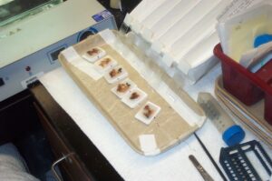
Tissues are processed for microscopic examination in a process developed more than a century ago. The tissue is preserved (most often using a formaldehyde solution; formalin). Then all the water is gradually removed by immersing the tissue in increasingly concentrated alcohols. The spaces where water once was are then filled with paraffin, a common and inexpensive wax.
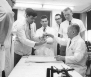
The paraffin is cooled and hardened into wax “blocks” so it can be sliced into very thin (3-4 micrometers; 1 micrometer is the same as 1/25,400 of an inch) “sections” which are placed on special glass “slides” which are then stained to display the nuclear and cytoplasmic details. Much of this processing is automatically done in specially designed machines. For decades, this processing was all done manually but, still, results were often ready by the end of the next day. When I began my residency, in 1965, the transition from manual to automated was in its early stages at our hospital—it was already the practice at other institution but our brilliant head of surgical pathology, Dr. Sadao Otani, was wary about trusting the vitally important tissue samples to machines—so my fellow residents and I learned much about handling tissues that today’s young pathologists may never know.
The stained slides are then studied by a pathologist—a physician with 4-5 or more years of study following
medical school graduation. A pathologist uses a microscope providing up to 400 times magnification. When blood and bone marrow (“smears”) are studied microscopically, a special objective lens enabling magnification to 1000 times is used. The pathologist typically dictates a report into the laboratory computer which he/she proofreads before completing (“signing out”). The report is then available to the patient’s physicians. “Deepers” means the pathologist felt he/she needed to look at another level of the “block.” All well and good, except that, again in the ideal setting, that can be accomplished within that first day after biopsy.
In that “ideal” setting, my biopsy report should have been ready at some time on Friday, March 15 or, at the latest, some time on Monday, March 18, two working days or four actual days after the biopsy was obtained.
Four days isn’t that long a time, you may say. But it is close to eons if you are waiting to learn the result of a stomach biopsy or a breast biopsy or a lymph node biopsy or, to get closer to my case, a possible skin cancer, such as melanoma.
Is there ever that “ideal” setting?
Of course.
A few years after I became Chairman of Pathology at Cedars-Sinai Medical Center, in Los Angeles, we asked the histotechnologists (the people who take care of all that tissue processing summarized above) if some, or all, would be willing to start their day at 5 AM or even 4 AM. Resoundingly, the answer was “yes.” They were more than willing to be adjust their schedules to be able to enjoy the sunny Southern California afternoons.
Consequently, slides for microscopy of biopsies were available to the residents and staff pathologists by 7 and 8 AM. In most instances, the residents examined them as soon as they received them and reviewed them with the staff pathologist before noon. The pathologist could dictate the diagnoses into the hospital computer system, add relevant images of the macroscopic and/or the microscopic appearances, and complete the case for the treating physician to see the diagnosis by early afternoon or earlier. Slides from bigger specimens, which are often less urgent and can benefit by slightly longer processing times, were usually available by 11 AM or early the next day. A less urgent approach was appropriate especially if a prior biopsy had been performed to establish the diagnosis and/or if the entire lesion was resected.
For biopsies, of course, this meant that phone calls could be made early about cases that might be urgent, as well as those with unexpected diagnoses, enabling other studies (X-rays, etc) to be ordered, treatment to begin and, most important, patient anxieties allayed. One of the first things our residents learned was that there was a living, concerned patient and/or family member associated with those biopsies, tissues or organs submitted for our studies and the sooner the patient and family learned the diagnosis, whether good or bad, the better.
When an important result was not available in this time (additional stains or other special studies needed) the pathologist generally called the clinician to inform him/her of the problem and, often, to provide an initial impression (e.g. benign or malignant, infectious or not infectious, etc).
I started writing this blog late in the afternoon on Thursday, March 21, the 7th day after my biopsy.
In my new career as a novelist and short story author, I am tempted to imagine the catastrophes that may have impacted on the timeliness of my results, although I know, just based on its gross appearance, that my lesion is benign.
It’s completely understandable that busy practitioners can’t look at every report as soon as it is generated. Usually, physicians set aside time at the end of the day or the beginning of the next day to review the wealth of ancillary data that relate to their patients.
On Friday afternoon, March 22, the 8th day after the biopsy was performed, I received a copy of the dermatopathologist’s report and a separate note from the dermatologist:
The spot came back from lab as a wart(1), which is a harmless overgrowth of skin thought to be caused by a virus called HPV. However, given the size and rapid growth of the spot, I question whether this is an accurate diagnosis and suggest full removal with additional pathology evaluation.
I immediately replied:
Thank you for your note. I was also surprised by the diagnosis. As you saw, the lesion has a definite cuff of normal skin, resembling keratoacanthoma or inverted papillary keratosis. Rarely, verruca vulgaris can also look like this. Of course, if the lesion has the typical eosinophilic inclusions, that resolves the question. Can you ask Dr. xx to send me copies of the images or recuts of the slides?
No matter what, I know it must be excised. I am not a Mohs (2) expert but this doesn’t seem to be the kind of lesion you treat with Mohs. What do you think?
Should I find a general surgeon to cut it out?
I am still astounded it took 8 days to get the diagnosis. I heard the whole world was made in 7 …
Best wishes.
Again, my note was answered by another assistant who did not respond to most of my comments and questions, even after I asked a second time.
I have subsequently learned that the usual turnaround for pathology specimens at this hospital is, indeed, 24-36 hours. Apparently, dermatopathology is not a part of pathology and its management, and processing standards, differ from those of the Department of Pathology.
I suppose the comment about the world being created in 7 days was a bit gratuitous …
I guess I was “venting my spleen” (3), something I don’t do very often.
* Initially, all diagnoses of tissue changes were rendered only on the basis of macroscopic (“gross”) examination. The microscopic, despite being invented in the 16th century, did not become widely used for medical purposes until the mid-19th century. Tissue processing for microscopic study did not become refined until after World War II and some pathologists were renowned for their macroscopic evaluations, which was refined at the University of Vienna. I was fortunate to have many teachers during my residency who studied in Vienna or were themselves taught in the Viennese tradition. Many factors contributed to the diminished reliance on macroscopic interpretations. In addition to the precision of microscopy becoming obvious and serving as the ‘gold standard’ for tissue diagnoses, techniques for preparing tissues for microscopy became refined and largely automated. This essay is not the place for expounding on this topic except to note that those who once learned the importance of careful gross examination know it is still important, whereas those who never learned don’t know. When I was a resident, my great teacher, Dr. Sadao Otani, was asked by a new
resident, who was working on a large specimen, “Dr. Otani. How many sections (samples for microscopic examination) should I take.” Otani responded, “The right one.” He didn’t mean that only one section should be taken (although sometimes that is enough). He meant that the selection of the section should follow a careful gross examination. If an area is bloody and necrotic, it generally won’t yield valuable information except for certain tumors, such as those in the ovary and testes, so it needn’t be sampled. In ovarian and testicular tumors, bloody and necrotic areas can be the most important to sample.
1 The diagnosis was “verruca vulgaris.” Verruca vulgaris is the term used for the common wart, a small, fleshy, painless bump on the skin or sometimes in the mouth and other places, caused by human papillomavirus (HPV). Characteristically, the cells of the growth have a distinctive, easily recognized accumulation (inclusion) indicative of the viral origin. Warts are common and almost always completely benign. They are typically flat or may have tiny projections, as mine does. As the name implies, the process can look “vulgar” and, for that reason, I have not included a photo of my lesion.
2 “Mohs microsurgery” is a technique developed in the 1930s but not popularized until the 1960s. It is used to remove the two most common types of skin cancer, basal cell carcinoma and squamous cell carcinoma. Its purpose is to minimize the injury to the surrounding skin while completely eradicating the tumor.
3 Vent one’s spleen. This phrase may date back to the 1600s, and refers to expressing one’s anger, as in “The community members tended to use town council meetings as a place where they could vent their spleen.” Vent is used in the sense of “air” and spleen represents “anger,” as a reminder that this organ was once considered to be the seat of ill humor and melancholy.


March 27, 2024 at 4:18 pm
Oh, my! I would go so far as to say that a dermatologist who does not have an “affinity for macroscopic evaluation” cannot possibly be a good dermatologist.
About 25 years ago, I took a very interesting class on measuring quality in the delivery of primary care. It was based on Avedis Donabedian’s “structure, process, outcome” model of measuring the quality of health care. One day, the professor, who believed that part of the job of primary care physicians was to coordinate specialty care, spoke about which physician specialists patients should be able to see without first seeing a primary care provider. She argued that elderly patients, who frequently have lots of benign skin lesions all over their bodies, should be able to go straight to dermatologists without first seeing their primary care providers.
What is a dermatologist who doesn’t have an affinity for macroscopic evaluation going to do? Biopsy every lesion on an elderly patient’s body? When you see a dermatologist instead of a primary care physician for a skin lesion, part of the value is that you can trust the former more than the latter to know which lesions need to be removed. That decision can only be made by a physician with an “affinity for macroscopic evaluation”.
March 27, 2024 at 5:44 pm
Hi Stephen,
I share your frustration. You should not be surprised because nowadays it is rare for anything to go smoothly. For some perverse reason the age of computers and high technology has also brought with it confusion and lack of efficiency. Whether it is the bank, supermarket, mail order medicine, postal service or any field in which thought and common sense are required f**k ups are the norm. Try not to vent your spleen. You may need it in the future.
Best regards,
Debby Troy Barcan
March 27, 2024 at 10:20 pm
quite a lesson, Steve…..the system you built in LA was indeed a model, and naturally i have speculated a bit regarding the hospital in NYC where this mismanagement took place. Glad it was in fact benign
March 28, 2024 at 12:35 am
I suspect your (our) age unfortunately makes reporting a pathogy report is sometimes on the “back burner”. Regardless , I also expect we will be reading your interesting tomes for many years to come.
March 28, 2024 at 6:59 am
As a person whose only medical experience is as a patient, I have grown accustomed to delay at every point in the medical treatment process (from just getting an appointment to aspects of treatment) which I assume is how physicians want it, possibly to reduce their own stress. If that sounds hypocritical, so be it. Often I just get to see a medical practitioner anyway. Like most patients, I just hope that my doctors are the more dedicated ones, but how could I tell? Perhaps by comparing my treatment in a very serious case….but I’d rather not HAVE a very serious case. It’s a quandary! Meanwhile, keep the blogs coming. Your well written insights are, as always, useful and entertaining.
March 28, 2024 at 8:45 am
Gosh, last I looked the world was created in six days, the seventh was a day of rest (Gen 2:1-2)
March 28, 2024 at 10:10 am
“ I heard the whole world was made in 7 …”
Steve, You are brutal! Ha ha. Perhaps there is a bit of Nathan Friedman in you! I have several LA friends who have bailed from the unwieldy, delayed care they get at UCLA and have moved over to a place called Cedars Sinai. Have you ever heard of that hospital? 🙂
April 7, 2024 at 6:09 pm
Very thought-provoking essay. Always appreciate your insight! Thank you for sharing your experience and perspective.
Amir